01 / DIAGNOSTIC EQUIPMENT
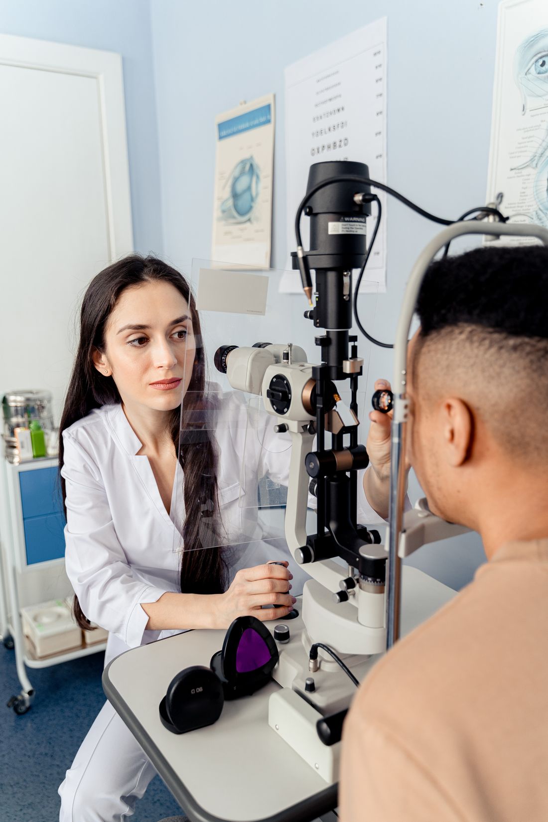
Slit-Lamp Biomicroscope
A binocular microscope with an adjustable beam of light, used to examine the cornea, iris and natural lens.

A closer look at instruments used in ophthalmic care.

From retinal imaging to refraction, different instruments answer different diagnostic questions. Explore how each technology is used before your visit.
A binocular microscope with an adjustable beam of light, used to examine the cornea, iris and natural lens.

Optical coherence tomography creates cross-sectional images of retinal layers and can assist with macular and optic-nerve evaluation.

Measures pressure inside the eye; this is one part of glaucoma evaluation, not a stand-alone diagnostic test.
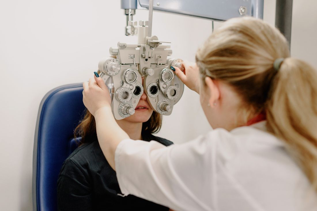
Interchangeable lenses help assess spectacle correction and guide a prescription for glasses.

Photographs the retina, including the optic disc and macula, supporting documentation of retinal findings.

A controlled visual-field test can detect areas of reduced peripheral or central sensitivity.
These real equipment photographs and diagnostic scans illustrate ophthalmic technologies; they are not photographs of Lensora's installed devices. The exact tests and machines available at the centre must be confirmed directly.
Not every patient needs every test. The choice of examinations depends on age, symptoms, medical history and clinical findings. A comprehensive eye examination is more than an equipment checklist.
How the tests work
Tell us what you need, and request a visit with the Lensora team.